
Ruth Schuster Feb 16 2016
MRI is the best way to scan the heart when trouble is suspected, but you can't move in the machine. With Naomi Chesler's stepper, the device sees your heart at work.
An MRI offers the best non-invasive snapshot of what's going on in your body. But because the coffin-like machine renders the body largely immobile, it's been useless in monitoring the heart at hard work - until now. A new invention allows patients to engage in exercise while being scanned, enabling doctors to detect heart trouble sooner and more efficiently than has been possible so far.
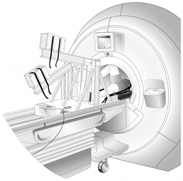
The concept is screamingly simple, once somebody had the idea – an exercise machine made out of special metal that can be used in proximity, or even inside, MRIs.
The device, a stepper designed by biomedical engineering professor Naomi Chesler and her team has two unusual features: You use it lying on your back, with your head and chest inside the MRI machine, and it's made of metals that magnets ignore.
MRI stands for "magnetic resonance imaging." It uses a powerful magnetic field to create images of the body’s internal organs and goes haywire in the proximity of ferromagnetic metals like iron. Any machine used around MRIs has to be nonferrous.
Also, the stepper is cheap, at less than $500 a pop, says Chesler, a professor at the University of Wisconsin and a visiting senior Fulbright scholar at Tel Aviv University, who developed the exercise machine with her team in the United States.
So far doctors have had to rely on echocardiography as a non-invasive imaging technique measuring heart function during exercise stress tests. MRI is preferable because it has higher spatial resolution, explains the doctor; with her device, it too can now measure the heart under stress.
Asymptomatic at rest
As people live longer, cardiovascular disease has become a leading cause of death worldwide. The earlier cardiovascular problems are diagnosed, the more can be done to help the patient. Chesler's breakthrough is to make an exercise machine that can be used while the MRI is scanning your secrets.
Why does cardiovascular data need to be collected during exercise, as opposed to the patient at rest, anyway? It's no great trick for the heart to function when you're doing nothing. The question is what it does when you exercise.
"Stress tests, including exercise stress tests (as opposed to pharmacological stress tests) provide clinically important additional information about cardiac function," explains Chesler. "For example, many patients are asymptomatic at rest but have angina with exercise."
The stepper her group created can stress the heart at a range of levels, from ambling out to get the mail, to going up stairs. Yes, even the meager exercise involved in going to get the mail can cause people with cardiovascular disease to lose their breath: "In that sense, it is 'exercise,'" Chesler tells Haaretz. "We define exercise as a measureable increase in cardiac output."
How does it work? You lie on the MRI bed, fit your feet into the pedal-like footholds and step as if walking on air while the MRI takes fast, high-resolution images of the heart and blood flow.
Using Chesler's machine, a few minutes at two or three different levels of exercise, plus measurements at rest, are enough for conclusions to be reached, she says.
The design of the machine is described in a paper "Low Cost Magnetic Resonance Imaging-Compatible Stepper Exercise Device for Use in Cardiac Stress Tests". Its use in measuring the stiffness of large arteries, which other researchers have shown can predict heart failure in patients with cardiovascular disease, is described in another paper, "Non-invasive measurement using cardiovascular magnetic resonance of changes in pulmonary artery stiffness with exercise".
While testing their machine's efficacy, Chesler's team also made a breakthrough in heart science, becoming the first to demonstrate that arterial stiffness increases in response to exercise in healthy human subjects (as opposed to healthy rat subjects).
"This is important because stiffness of the main pulmonary artery (at rest) is a strong predictor of mortality in patients with pulmonary arterial hypertension," she says. "Our study helps us understand the factors that affect main pulmonary artery stiffness and contributes to its utility as a clinical metric."
So far the device has been tested on healthy people and people with pulmonary arterial hypertension, and the team is confident it will help with diagnostics of known or suspected cardiovascular disease. "We know it 'works' because we can measure main pulmonary artery stiffness at rest and during exercise with our device, using the MRI," Chesler explains.
Ruth Schuster
Haaretz Correspondent
read more: http://www.haaretz.com/israel-news/science/1.703368